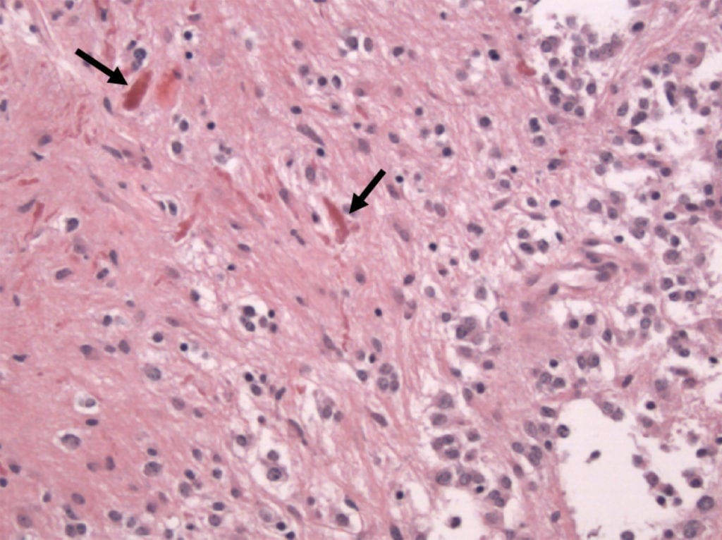
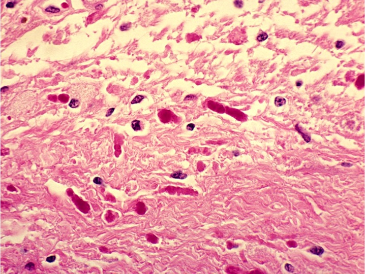
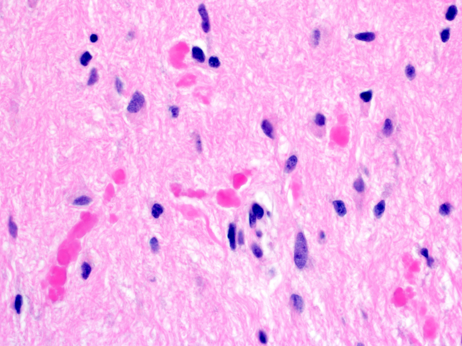
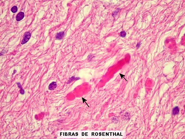
The neoplasm may also be solid. Rosenthal fibers (rfs) are cytoplasmic, proteinaceous aggregates. They are the pathognomonic feature of the astrocyte pathology in alexander disease (axd), a neurodegenerative disorder caused by heterozygous mutations in the gfap gene, encoding glial fibrillary acidic protein (gfap).
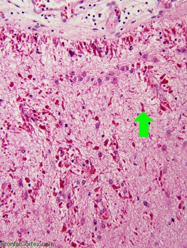
Although rfs have been known for many years their origin and. The presence of eosinophilic rosenthal fibers is a characteristic feature and hyalinization of blood vessels Pilocytic astrocytoma (pca), previously known as cystic cerebellar astrocytoma or juvenile pilocytic astrocytoma, was first described in 1931 by harvey cushing, based on a case series of cerebellar astrocytomas;
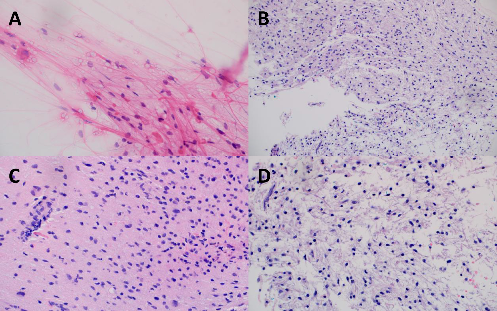
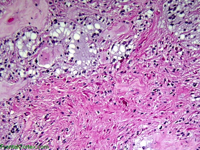
Histopathological aspects of pilocytic astrocytoma. All microphotographs represent h&e stain. Note the vasculature, with typical endothelia (200x).
Pilomyxoid astrocytomas (pmas) were first officially described in 2007. Since then, intermediate pilomyxoid tumors with histopathological features typical of both pmas and pilocytic astrocytomas (pas) have been described. However, we found evidence of tumors that are histologically like pmas but contain rare rosenthal fibers, which have been.
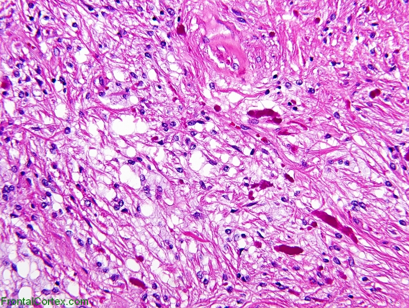
Rosenthal fibers. 10 the purpose of this report is to review the nature and significance of rosenthal fibers in the context of glial fibers, alexander disease, and such tumors as pilocytic astrocytomas. Histology of pilocytic astrocytomas with prominent rosenthal fibers image and description are from the afip atlas of tumor pathology: Government work which may be used without.
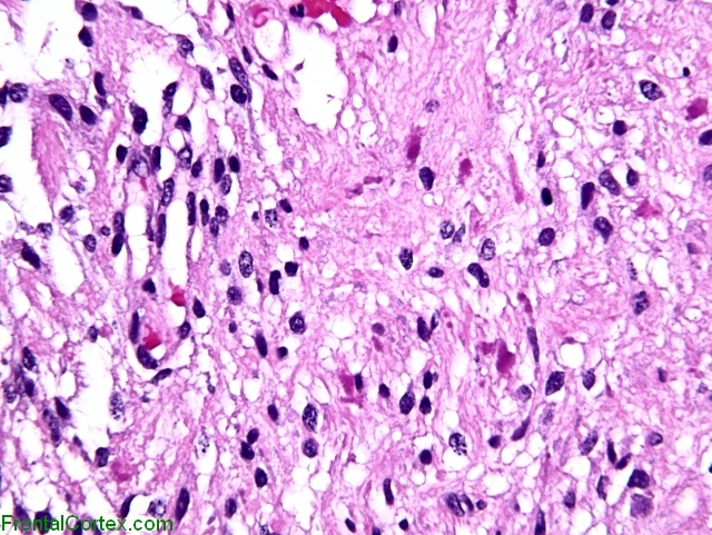
Rosenthal fibers high quality pathology images of neuropath, glial tumors, pilocytic astrocytoma. Another useful diagnostic feature is pas+ve eosinophilic granular bodies. Slide 8 of 19.
Immunohistochemically we investigated rosenthal fibers (rfs) on specimen surgically removed from patients with glioma (three cerebellar astrocytomas, three optic gliomas, two spinal cord astrocytomas, one spinal ganglioglioma). Pathological diagnoses were pilocytic astrocytoma, fibrillary astrocytoma, and ganglioglioma. The histology of pilocytic astrocytoma is biphasic with areas of bipolar cells and rosenthal fibers and multipolar cells with eosinophilic granular bodies and microcysts.
Rare mitotic figures, microvascular proliferation, necrosis, and invasion of meninges can be. Classically in the cerebellum in children; Most common glioma in children.
[1] the optic glioma is associated with neurofibromatosis 1. Rare variants include pilomyxoid astrocytoma and anaplastic pilocytic astrocytoma. In summary, there are pilomyxoid tumors that are similar to pmas histologically but also have rare rosenthal fibers.
Its presence is associated with either pilocytic astrocytoma (more common) or alexander's disease (a rare leukodystrophy). They are also seen in the context of fucosidosis. Pilocytic astrocytoma is the most common primitive tumor found in pediatrics.
Studies of more such cases are needed for clarification of such tumors’ clinical features. Keywords astrocytoma pilomyxoid pilocytic rosenthal fibers tumor introduction in 1999, tihan et al. Intermediate filaments and gfap.
A deeper understanding of rosenthal fibers is greatly facilitated by an appreciation of the cytoskeletal architecture of the typical astrocyte. Rosenthal fibers and eosinophilic granular bodies (egbs) activating genetic alterations in components of mapk pathway (most frequently braf fusions) (nat genet.


.jpg)