Symptoms and ecg findings in both pericarditis and stemi look similar. It is commonly caused by an acute occlusion (the blockage of a blood vessel or hollow organ) of a coronary blood vessel secondary to acute plaque rupture (an area of fibrous cap disruption) and thrombosis. Difference between pericarditis and.
C) liberty hospital connected to you. 4. 4 stemi vs pericarditis; Differentiating stemi from pericarditis.
A randomized trial of colchicine for acute pericarditis. n engl j. Pericardial effusion (larger than trivial) there are no studies that have determined clear diagnostic criteria. One of the biggest pitfalls in the diagnosis and treatment of pericarditis is misinterpretation of the ecg.
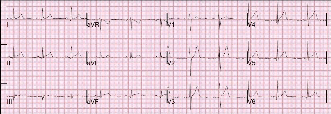
Presence of pr depression was the best discriminator pointing toward a diagnosis of pericarditis. However, pr depression was present in 12% of stemis, and spodick’s sign was present in 5% of stemi patients. In fact, every ecg finding had overlap between the two diagnoses.
To diagnose pericarditis we need a minimum of 2 of the following 4 criteria: 1 typical chest pain. 3 typical ecg changes.
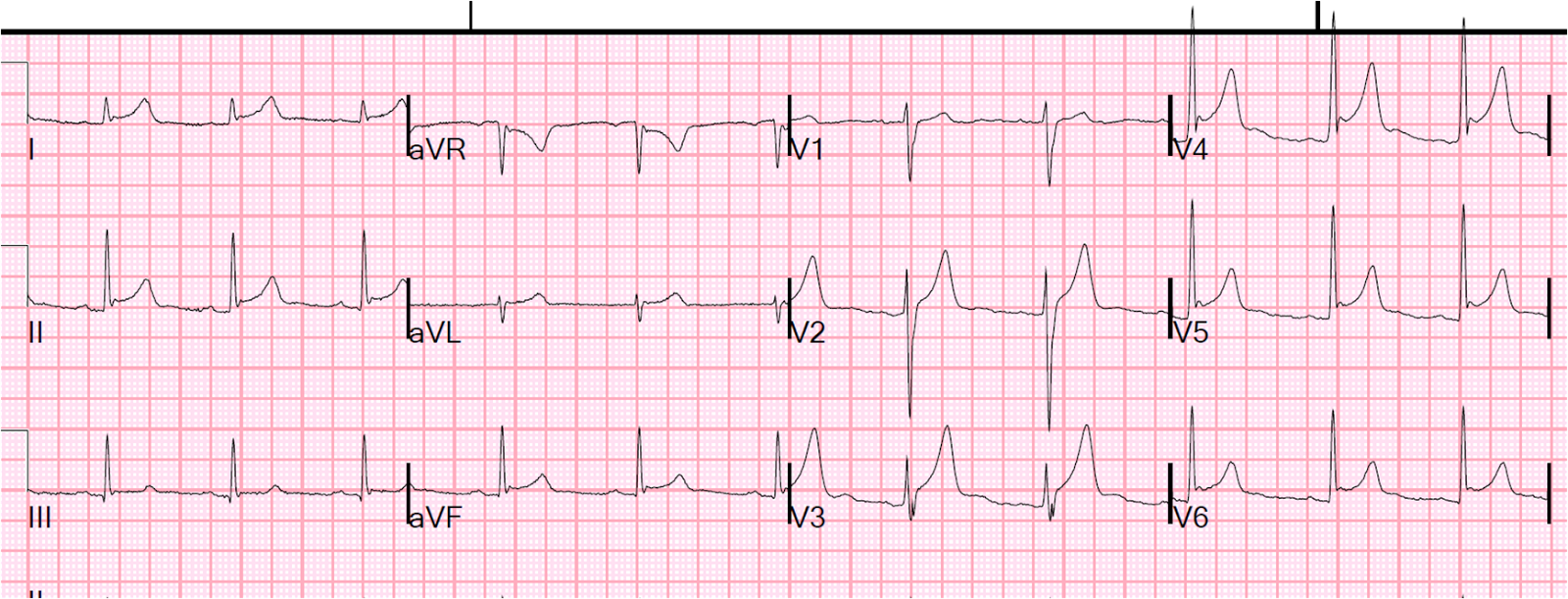
Has q waves, no reciprocal changes, similar to old ecg. If you cannot explain st elevation clinically, consider stat echo in the ed to evaluate for other causes. Lv aneurysm causes persistent st elevation due to wall motion defect.
Lv aneurysms are clinically important high risk for arrhythmia, thrombus formation. So what is a stemi? The st segment of an ekg will be elevated by more than 3 mm in the precordial leads and by more than 1 mm in the limb leads.
Further, in the precordial leads, the shape should be convex up (like a tombstone). The shape matters more than the amount of elevation in some cases. You diagnose pericarditis at your (or your patient's) peril.
Pericarditis does not have reciprocal st depression, not in either lead avl or in v2. This ecg is very specific for stemi. Even when patients are asymptomatic, or apparently so, they can have life threatening mi.
If the ecg has specific signs of mi, as this one does, then do. This content is for members only. Get a full year access for only $26!
Acute pericarditis is an inflammation of the pericardium. This inflammation causes ekg changes that have typically evolved sequentially through 4 stages 1. Witting md et al.
Evaluation of spodick’s sign and other electrocardiographic findings as indicators of stemi and pericarditis. Pericarditis is a condition that affects the pericardium, a sac that surrounds the heart. The pericardium can become inflamed (called pericarditis), which can lead to chest pain (stemi) and shortness of breath (respiratory distress).
Pericarditis is most common in people over 50 years old, but it can also occur in younger people. Pericarditis is the inflammation of the pericardium while stemi is a very dangerous type of heart attack where a major artery is completely blocked. Most of the patients with pericarditis experience sharp, piercing pain in the left side or center of the chest.
Stemi’s signs and symptoms include chest pain or discomfort, dizziness, nausea. Refer to panel 1 for all ecg criteria for stemi. Ecg criteria for the diagnosis of acute stemi.
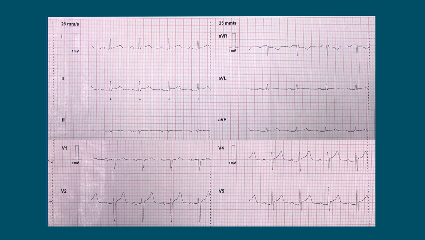
New st segment elevations in at least two anatomically contiguous leads: The diagnostic yield of. However, an unusual ecg presentation of the simultaneous occurrence of the two conditions has not been reported previously.
Acute pericarditis tends to affect younger individuals. The most common cause of pericarditis is infections, which is why many patients may report symptoms consistent with viral infections (particularly in the preceding days). The ecg in acute pericarditis (myocarditis) the ecg is highly effective in differentiating pericarditis from stemi.